


Diagnostic services for bumps and growths involve running tests or taking a biopsy to determine their underlying cause. A biopsy involves taking a sample from the affected area and testing it in a lab to look for signs of disease. Many bumps and growths found in or below the skin turn out to be harmless. Patients who are concerned about these bumps and growths should watch for changes or symptoms that could indicate an underlying condition that requires treatment.
What Causes Skin Growths?
Genetics
Some skin is simply more prone to warts and growths than others. This is due to genetic factors related to your skin type, as well as genetic predispositions to certain conditions that cause growths, which are not uncommon.

Underlying Conditions
Conditions like allergic eczema, warts, molluscum contagiosum, scabies, and sebaceous cysts can range from mild to severe. Some require minimal care, while others can be life-threatening without proper treatment.

External Factors
External factors like insect stings, splinters, and illness can cause temporary growths to appear on the skin. Oftentimes these itch or sting and are symptomatic of something more.
Underlying Conditions
Bumps and growths may go away if you treat the underlying problem, though there are alternative options for patients if this doesn’t work. Some cysts, for example, can be drained, but other lumps and growths may require surgical removal.
Prevention
Proper skincare and management reduce the risk of infections and other problems. People should be aware that skin cancer and other severe disorders of the skin that might result in growths and bumps call for prompt medical attention and assessment. This significantly reduces the possibility of future complications.
Our Process
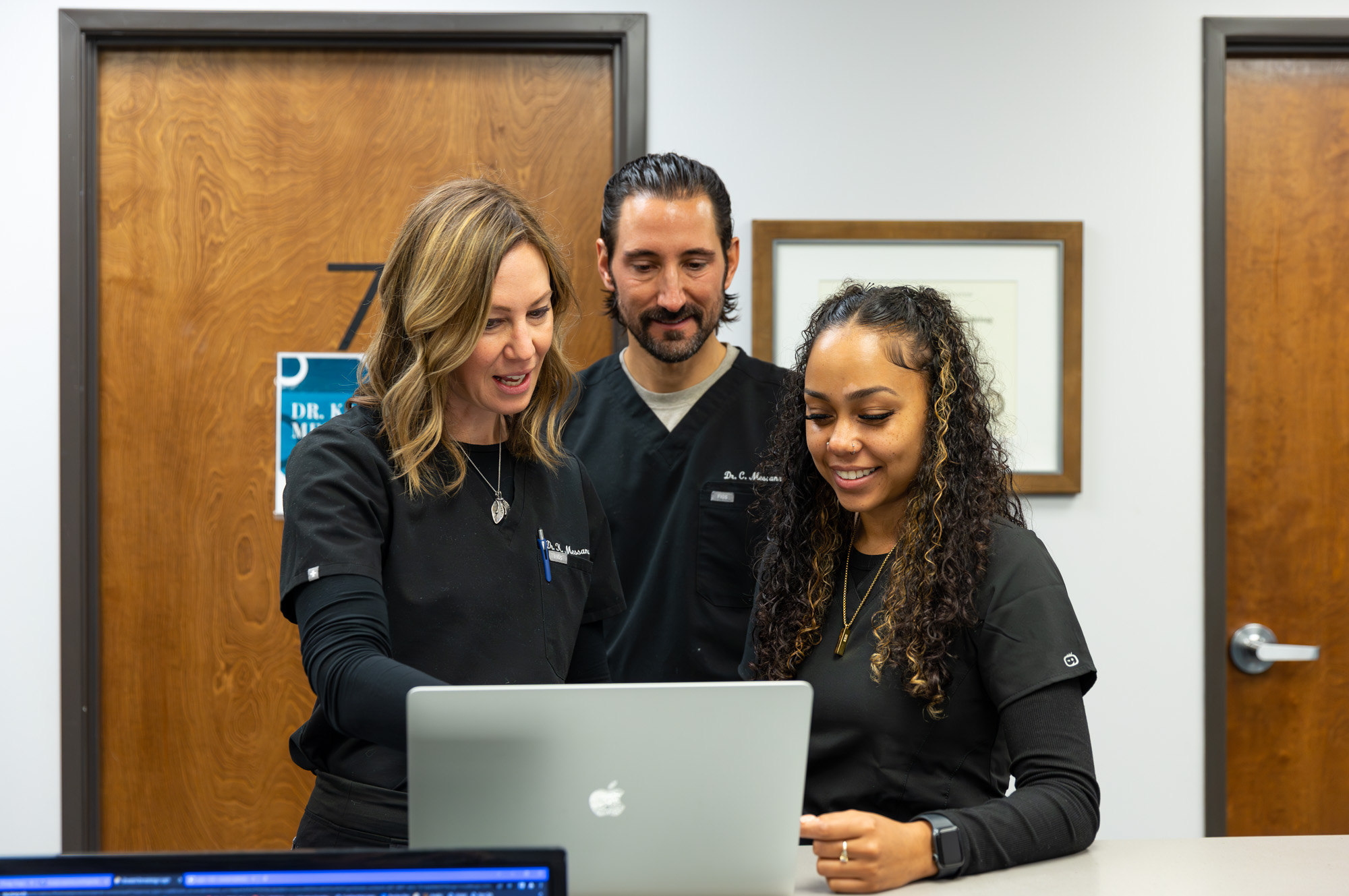
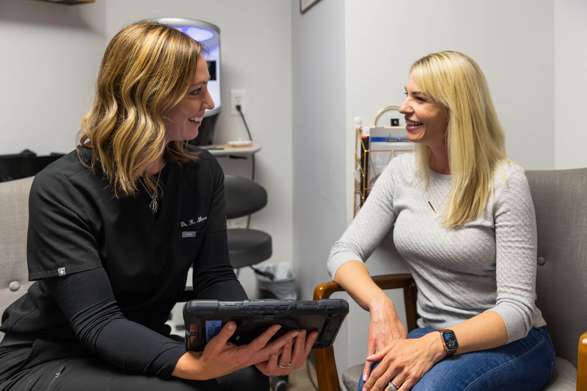
Consultation
At Elevated Dermatology, we know every patient is unique. We start with a thorough consultation to explore all your concerns, goals, and options for skin growth removal in Parker, CO.
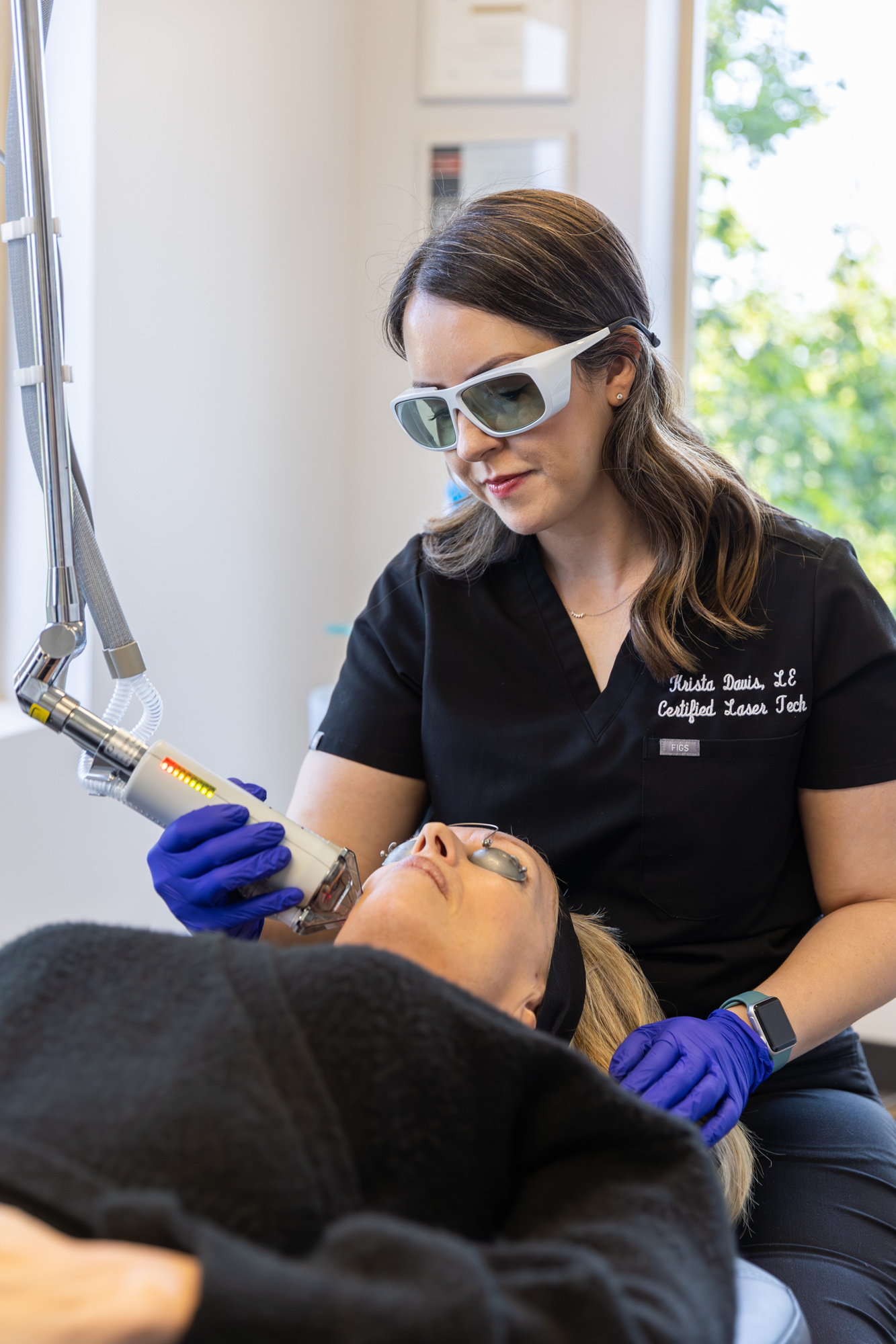
Treatment
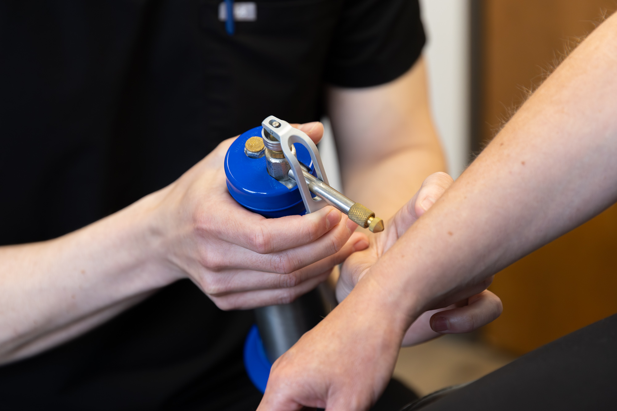
First, we'll assess your symptoms and look at possible conditions that could result in your particular concern. Once we discover the root cause, we treat it accordingly with things like topical creams, drainage, or an excision.
Results
The results vary based on the condition being treated; however, at Elevated Dermatology Skin Cancer Surgery Center, we strive to improve your quality of life and your skin.


Frequently Asked Questions About Bumps and Growths
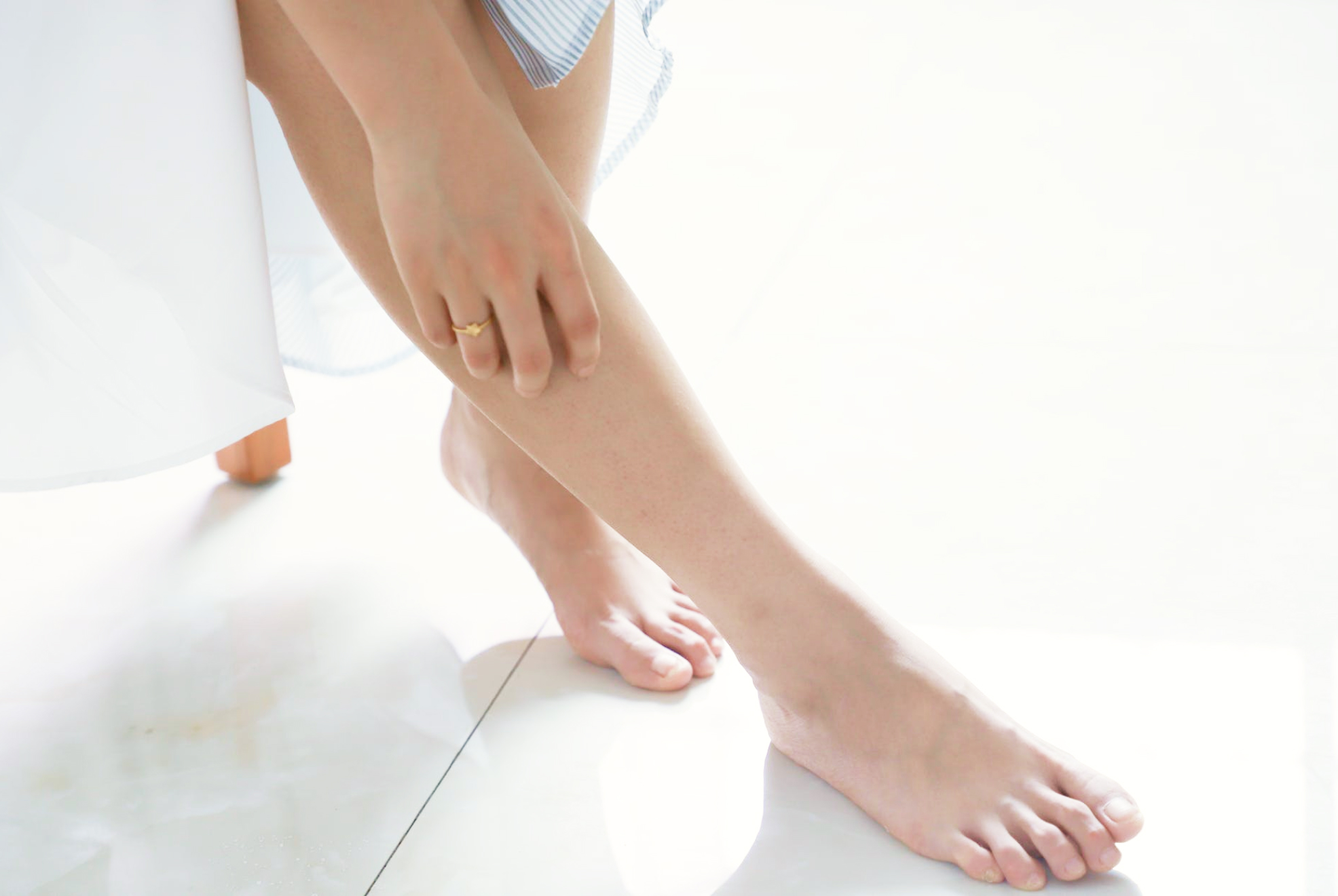
Those with bumps and growths that change appearance should have them checked. Even when non-dermatologists consider bumps and growths benign or harmless, it’s important to have dermatologists check them to look for signs of cancer or other conditions that need treatment. Patients should watch for changes in size and shape or the presence of symptoms, such as itching, discomfort, bleeding, or burning.
First and foremost, we examine your concerns and seek to provide answers. A number of underlying conditions and problems can cause bumps and skin growth. Once we establish what issue could be causing new bumps, growths, or changes, we can take appropriate action.